Exploring the Formation and Effects of Epiretinal Membrane

Exploring the Formation and Effects of Epiretinal Membrane

Exploring the Formation and Effects of Epiretinal Membrane

Exploring the Formation and Effects of Epiretinal Membrane

Have you ever experienced a slight distortion in your vision, almost as if you're looking through a piece of cellophane? That thin layer that could be causing this visual disturbance is known as an epiretinal membrane. Your eyes are complex organs with various parts working harmoniously to provide you with clear vision. However, sometimes things can go awry, and an epiretinal membrane is one such condition that may affect your eyesight.
What is an Epiretinal Membrane?
An epiretinal membrane is a thin, fibrous layer that forms on the surface of the retina, specifically on the macula, which is responsible for your central vision. This layer is also sometimes referred to as a macular pucker or cellophane maculopathy. It's a translucent sheath that develops because of cellular changes in your eye. Over time, this membrane can contract and cause the underlying retina to wrinkle or pucker, which in turn affects your vision.
The exact mechanism behind the formation of an epiretinal membrane is complex. It often begins when the vitreous, the gel-like substance filling your eye, starts to shrink and separate from the retina as part of the natural aging process. This can lead to the release of cells that migrate and proliferate on the retinal surface, forming the epiretinal membrane. In some cases, the membrane may develop following eye surgery, injury, or because of other eye conditions.
An epiretinal membrane is composed of collagen, fibrous cells and, occasionally, blood vessels. The density and contractile properties of the membrane can vary which determines the severity of the visual disturbance. In many cases, the membrane is thin and may not cause significant symptoms. However, in other instances, the contraction can be severe enough to distort your vision and necessitate intervention.
The Causes of an Epiretinal Membrane
Various eye conditions can lead to the development of a secondary epiretinal membrane. For instance, individuals with a history of retinal detachment, uveitis (eye inflammation), or vein occlusions in the eye are at greater risk. Additionally, those with proliferative diabetic retinopathy may experience abnormal growth of blood vessels, which can contribute to membrane formation.
Age is a significant risk factor for the development of an epiretinal membrane, with most cases occurring in individuals over the age of 50. However, it's not limited to the elderly; traumatic eye injuries and prior ocular surgeries, such as cataract extraction, can also increase the likelihood of developing an epiretinal membrane. Genetics may play a role, although this relationship is not yet fully understood.
Identifying the Symptoms of Epiretinal Membrane
The symptoms of an epiretinal membrane can be quite subtle, particularly in the early stages. You might notice a mild distortion in your vision, where straight lines appear wavy or bent. Difficulty reading fine print and experiencing blurred or double vision in one eye are common complaints. The changes in your vision can be gradual, making them easy to overlook initially.
For some individuals, the symptoms of an epiretinal membrane may remain stable and not worsen significantly over time. In others, the membrane can continue to contract, leading to progressive visual distortion and, in severe cases, a decrease in central vision. It's important to monitor any changes in your vision, no matter how minor they may seem, and report them to your eye care professional.
Diagnosing Epiretinal Membrane
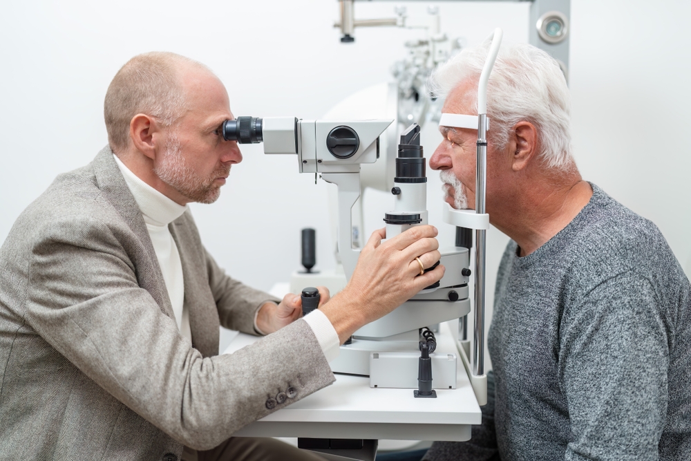
When you visit an ophthalmologist with concerns about your vision, they will perform a comprehensive eye exam. This includes checking your visual acuity, examining the front and back of your eye, and assessing your retina's health. The ophthalmologist will look for signs of an epiretinal membrane, such as wrinkling on the retinal surface, using specialized equipment.
To confirm the diagnosis of an epiretinal membrane and assess its impact on your retina, your ophthalmologist may utilize advanced diagnostic tools. Optical coherence tomography (OCT) is a non-invasive imaging test that provides detailed cross-sectional images of your retina, allowing the doctor to visualize the membrane and any associated retinal changes. Fundus photography and fluorescein angiography may also be employed to gain further insight into your eye's condition.
Depending on the severity of your symptoms and the impact on your vision, the ophthalmologist will discuss treatment options with you. In many cases, observation is the recommended course of action, especially if the symptoms are mild. However, if the epiretinal membrane significantly affects your quality of life, surgical intervention, such as a vitrectomy with membrane peeling, may be considered. Your ophthalmologist will guide you through the decision-making process, considering your individual needs and lifestyle.
Conclusion
An epiretinal membrane can be a daunting prospect, but with the right knowledge and care, it can be managed effectively. Don't let changes in your vision go unchecked. Schedule an appointment with your ophthalmologist today and take the necessary steps to protect your eyesight.
For more information on an epiretinal membrane and treatment options, contact our professionals at Gulf Coast Retina Center in our Sarasota or Venice, Florida, office. Be seen today or call (941) 312-2769 to schedule an appointment today.

All Eye
Care Services

Keep
In Touch
contact information
office hours
- Monday:8:00 AM - 5:00 PM
- Tuesday:8:00 AM - 5:00 PM
- Wednesday:8:00 AM - 5:00 PM
- Thursday:8:00 AM - 5:00 PM
- Friday:8:00 AM - 5:00 PM
- Saturday:Closed
- Sunday:Closed
© 2026 Gulf Coast Retina Center. All Rights Reserved.
Powered by: